Rewriting the Rules of Slumber
For nearly a century, the scientific consensus on sleep has been deeply anchored in a hierarchical view: ancient, deep-brain structures hold the keys to our wakefulness and rest, while the cerebral cortex—the brain's seat of higher cognition—merely tags along for the ride. However, a transformative study published in the journal Nature, led by Dr. Renata Batista-Brito, suggests the cortex is far more than a passive passenger. Researchers have uncovered an exceptionally rare population of neurons that may hold the power to actively orchestrate the onset of sleep.
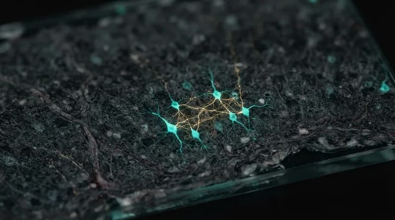
These specialized cells, known as Sst-Chodl neurons, function as long-range inhibitory interneurons. While most inhibitory neurons limit their communication to local neighborhood cells, Sst-Chodl neurons project signals across expansive regions of the neocortex. This unique anatomical configuration allows them to exert a sweeping influence, coordinating brain-wide activity that was previously thought to be driven exclusively from the brain's interior.
The Anatomy of a Sleeper Cell
Sst-Chodl neurons are a biological enigma, accounting for a mere 0.2% of the cortical neuronal population. Despite their scarcity, their impact is disproportionately profound. During periods of wakefulness, these neurons remain largely dormant. However, as an organism transitions into a drowsy state, these cells spring into action, firing in alignment with the slow, synchronized brain rhythms that characterize non-rapid eye movement (NREM) sleep.
The research team utilized optogenetic and electrical stimulation to test the functional capacity of these cells in mice. When the researchers artificially activated the Sst-Chodl neurons, the result was a nearly immediate shift in the cortex's electrical signature. The brain waves slowed down and became synchronized, creating a state that mimicked natural sleep. Furthermore, this activation was sufficient to drive mice into a resting state more quickly and increase their total sleep duration, proving these cells serve as a functional switch for sleep regulation.
Why It Matters
- Challenging Dogma: The study shifts the narrative from the cortex being a passive observer to an active participant in sleep initiation.
- Evolutionary Significance: These neurons have been preserved across hundreds of millions of years of evolution, appearing in species ranging from amphibians to humans, suggesting a critical role for survival.
- New Diagnostic Frontiers: Disruptions in sleep are common in conditions like Alzheimer’s disease and autism; this discovery provides a specific neurological target for future research into sleep-related pathologies.
Future Implications for Sleep Medicine
The discovery of Sst-Chodl neurons opens a critical door for neurology. Because these cells have been preserved through vast evolutionary timelines, it is highly probable that they perform the same fundamental, regulatory function in humans. One of the most compelling avenues for future investigation is the "sleep pressure" hypothesis. Researchers are now exploring whether these neurons act as biological sensors, monitoring the accumulation of sleep-inducing signals and eventually triggering the cortex to "switch off" once the need for rest becomes critical.
By identifying a specific circuit involved in the transition to sleep, scientists now have a concrete biological target to explore how sleep breaks down in disease states. This could lead to a deeper understanding of why individuals with certain neurodevelopmental or psychiatric disorders struggle to maintain healthy sleep cycles, potentially pointing toward novel, circuit-based therapeutic interventions that target the cortex directly rather than relying on systemic sedatives.