A Prehistoric Medical Record
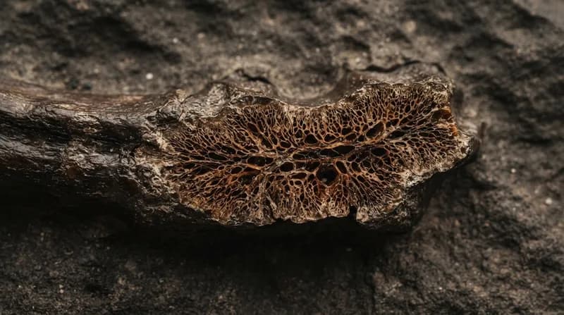
In a discovery that feels more like a scene from a science fiction film than a paleontological study, researchers have unearthed a stunning biological secret hidden within the bones of 'Scotty,' the largest Tyrannosaurus rex skeleton ever discovered. While examining a fractured rib from the massive predator, scientists identified a 66-million-year-old network of mineralized blood vessels that were actively forming as part of a healing process when the animal died. This extraordinary find offers a rare, high-resolution snapshot of dinosaur physiology that has remained protected within the fossilized bone for tens of millions of years.
Ordinarily, soft tissues such as blood vessels decompose long before the fossilization process can occur. However, the unique conditions of a salty marsh environment where Scotty’s remains were discovered acted as a geological time capsule. When the rib fractured, iron-rich blood rushed to the injury site, triggering the growth of new vessels to mend the damage. Because the dinosaur succumbed to its fate before the healing was complete, this microscopic medical record was preserved, providing scientists with an unprecedented view into the trauma and recovery of Earth’s most formidable predator.
The Power of Neutron and X-Ray Imaging
To analyze the fossil without risking damage, the research team—led by experts from the University of Regina and the Oak Ridge National Laboratory (ORNL)—employed a sophisticated suite of non-invasive imaging technologies. While standard X-ray methods provided a base-level understanding, the researchers leveraged the Department of Energy's specialized neutron facilities to probe the dense bone structure. Neutron imaging is uniquely suited for this task because, unlike X-rays, neutrons interact strongly with light elements like hydrogen, making them exceptionally effective at highlighting soft tissue remains and subtle chemical differences within a specimen.
The team utilized two state-of-the-art instruments at ORNL: the Multimodal Advanced Radiography Station (MARS) and the Virtual Environment for Neutron Sciences (VENUS). The MARS instrument, which generates 'cold' neutrons, proved vital for capturing high-resolution signatures of soft tissue. Meanwhile, the VENUS instrument provided high-energy neutrons capable of penetrating the thick, dense bone of the T. rex to construct detailed 3D internal maps. This multimodal approach allowed the researchers to corroborate earlier findings from synchrotron radiation techniques and visualize the fossilized vessel network in full three-dimensional detail for the first time.
Why It Matters
- Non-Invasive Discovery: By avoiding physical dissection, the researchers have preserved the integrity of the fossil while gaining data that was previously thought to be lost to time.
- New Diagnostic Potential: The application of neutron imaging to paleontology establishes a new protocol for studying the pathology of ancient life, allowing scientists to compare how injuries healed in extinct species versus modern animals.
- Technological Versatility: While neutron sources are typically used for materials science or battery testing, this research demonstrates their immense value in uncovering the biological mysteries hidden within our planet's fossil record.
Looking Toward the Future of Fossil Analysis
The success of the imaging campaign at ORNL has opened a new frontier in paleontology. Researchers are already looking beyond this single rib, with plans to apply these same neutron imaging techniques to a vast array of fossils currently sitting in museum collections. Many of these specimens likely hold their own 'hidden' secrets—from preserved scales to ancient amber inclusions—that have been inaccessible due to the limitations of traditional inspection methods.
By combining high-energy neutron beams with advanced data processing, the scientific community is entering an era where fossils can be treated as dynamic biological datasets rather than static stones. As the team continues to study the data from Scotty’s rib, they hope to map out larger trends in dinosaur health and injury patterns. This work not only refines our understanding of how individual dinosaurs lived and survived, but it also provides a clearer picture of the environmental pressures and evolutionary challenges faced by species during the final stages of the Cretaceous period.