A New Frontier in Early Oncology
In a major development for dermatological science, researchers at the Friedrich-Alexander-Universität Erlangen-Nürnberg (FAU) have successfully demonstrated a method to identify basal cell carcinoma—the most common form of skin cancer—well before physical symptoms manifest on the surface of the skin. Led by Dr. Moritz Ronicke at the Department of Dermatology at Uniklinikum Erlangen, the team has utilized advanced imaging technology paired with artificial intelligence to detect tumors in high-risk patients who show no visible signs of malignancy.
Basal cell carcinoma is particularly dangerous because of its tendency to spread locally and destroy surrounding tissue, often resulting in significant damage to sensitive facial areas like the nose or eyes. By catching the disease at this subclinical stage, medical professionals can pivot away from invasive surgeries toward more conservative, less painful treatments, such as targeted topical creams.
The Technology: Line-Field Confocal Optical Coherence Tomography (LC-OCT)
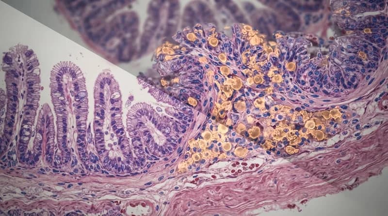
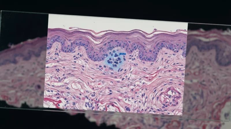
The core of this breakthrough lies in Line-Field Confocal Optical Coherence Tomography, or LC-OCT. This sophisticated imaging technique merges two powerful diagnostic modalities: optical coherence tomography, which maps the depth and structural extent of a tumor, and confocal microscopy, which captures high-fidelity images at the cellular level. This hybrid approach enables clinicians to generate three-dimensional, real-time visualizations of skin tissue with a resolution down to the micrometer level—a scale where even the finest human hair appears massive by comparison.
The Role of Artificial Intelligence
While the hardware provides the high-definition visuals, artificial intelligence serves as the analytical engine that accelerates the diagnostic process. The AI system processes the LC-OCT data in real time, assigning a color-coded probability value to the scanned areas that indicates the likelihood of basal cell carcinoma. This instantaneous feedback allows physicians to pinpoint suspicious regions during a routine examination that would otherwise remain undetected.
It is important to note that the AI acts as a sophisticated diagnostic assistant rather than an autonomous decision-maker; the final medical diagnosis remains firmly in the hands of the physician. The system is designed to act as a high-precision filter, highlighting areas that require immediate attention or monitoring.
Why It Matters
- Non-Invasive Potential: Early detection allows for the use of pharmaceutical creams instead of physical excision, significantly improving patient comfort and recovery.
- Preventing Disfigurement: By identifying tumors before they grow, doctors can prevent the tissue destruction that frequently necessitates complex facial reconstruction.
- Scalability: While the current method requires further data to assess sensitivity and speed, the integration of AI-assisted scanning could eventually become a standard preventative measure for high-risk populations.
Looking toward the future, the research team is focused on expanding the use of these 'subscans' to make them a routine part of clinical dermatological practice. Although further studies are required to refine the process and handle the current time-intensive requirements of the screening, this feasibility study published in JAMA Dermatology marks a pivotal shift in how we perceive the timeline of cancer diagnosis—moving from reactive treatment to proactive detection.