A Paradigm Shift in Neuroscience
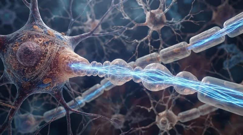
For over a hundred years, students and researchers alike have relied on a singular image of the brain’s microscopic wiring: axons depicted as smooth, consistent tubes carrying electrical impulses. However, a landmark study from Johns Hopkins Medicine has fundamentally challenged this century-old consensus. By utilizing high-pressure freezing electron microscopy—a technique that preserves cellular structures in a near-native state—scientists discovered that axons naturally feature repeating, pearl-like bulges along their length.
These structures, dubbed "non-synaptic varicosities," are not merely remnants of cellular damage or degenerative conditions like Parkinson’s disease, where similar swelling is often observed. Instead, these delicate "pearls" appear to be a standard, functional feature of healthy axons. This discovery forces a re-evaluation of how neuroscientists conceptualize the physical pathways that facilitate learning, memory, and cognitive function.
The Mechanics of Signal Speed
The research, published in Nature Neuroscience, suggests that this "pearls-on-a-string" architecture plays a direct role in regulating the speed of neural communication. By building mathematical models and testing them against mouse neurons, the team discovered that these swellings serve as more than just structural quirks. They act as dynamic components of the axon’s membrane, influenced by physical factors such as cholesterol levels and osmotic pressure.
The study found that neural activity itself can reshape these pearls. Following high-frequency stimulation, the varicosities were observed to stretch and widen, which directly influenced the speed at which electrical signals traversed the axon. By altering the diameter of these narrow channels, the brain may effectively modulate traffic flow, preventing signal congestion. The removal of cholesterol, for instance, altered the membrane's fluidity and significantly degraded the efficiency of these electrical messages, highlighting the role of membrane physics in brain health.
Why It Matters
- Rewriting Anatomy: The finding necessitates an update to biological textbooks, which have long simplified the geometry of nerve cells.
- Dynamic Signaling: It proves that axons are not fixed, static cables, but rather adaptable structures that physically respond to brain activity.
- Disease Distinction: Understanding the difference between these healthy, functional "pearls" and the pathological swelling associated with neurodegenerative diseases could open new avenues for diagnosing and treating neurological disorders.
- Human Validation: Subsequent research published in Neuron has confirmed similar structures in human cortical tissue, suggesting that this mechanism is a fundamental property of the human brain.
Future Implications
The transition from a static model of axons to a dynamic, physical understanding of membrane geometry is a significant leap for neurobiology. By recognizing that these axons can morph in response to electrical input, scientists now have a more complex map of how the brain regulates its own bandwidth. The next phase of this research, supported by ongoing grants from the National Institute of Mental Health, will focus on how these physical properties interface with chemical signals. As imaging technology continues to evolve, the distinction between functional structural changes and those signaling a decline in brain health will be vital for future clinical interventions.