A New Frontier in Imaging Technology
In a major development for material and biological sciences, a collaborative team of researchers across Austria—led by experts at TU Wien—is constructing a revolutionary imaging system that bridges the gap between electron microscopy and quantum computing. While conventional electron microscopes have long provided us with the ability to observe the atomic world, they rely on a relatively brute-force approach: bombarding samples with massive quantities of electrons to build an image. This process, while effective for inorganic materials, often destroys delicate biological specimens like proteins before a clear picture can even be captured.
The new project aims to move beyond simple electron counting. By integrating an ion-based quantum computer directly into the microscope's architecture, scientists hope to exploit the hidden quantum information carried by every electron. Instead of merely treating electrons as individual particles to be counted, the researchers are developing a system that processes the quantum states of these electrons, turning what would normally be discarded as random noise into high-fidelity data signals.
How the Quantum-Electron Hybrid Works
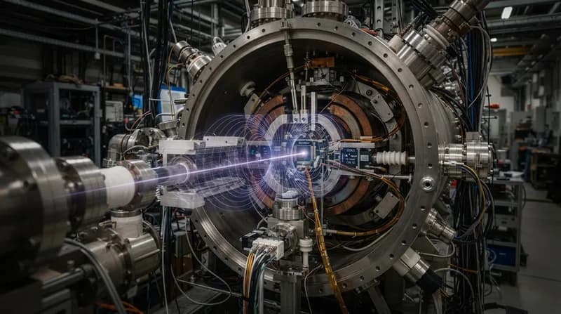
The innovation centers on the phenomenon of quantum entanglement. As electrons travel through the microscope and interact with a specimen, they are funneled through a region containing trapped ions. These ions serve as the building blocks of a quantum computer. As the electrons pass by, they become entangled with these ions, effectively sharing a quantum state. This allows information about the electron—which would otherwise be lost in a standard imaging setup—to be stored and manipulated within the quantum memory of the trapped-ion system.
Once this information is captured, the system performs specialized quantum-computing algorithms designed to aggregate data from multiple electrons. By processing these interactions, the microscope can reconstruct a high-resolution image using only a fraction of the electron dose required by current technology. This advancement effectively overcomes the statistical boundaries that have constrained imaging resolution for decades, providing a much clearer window into the structure of sensitive materials without the risk of high-energy degradation.
Why It Matters
- Preservation: Dramatically lower electron doses mean that biological samples can be imaged in their natural, delicate states without radiation damage.
- Signal Efficiency: The quantum integration extracts useful signals from background noise, potentially revealing structures that were previously invisible.
- Technological Convergence: This project represents a successful fusion of two cutting-edge fields: high-resolution microscopy and quantum information science.
From Mathematical Theory to Experimental Reality
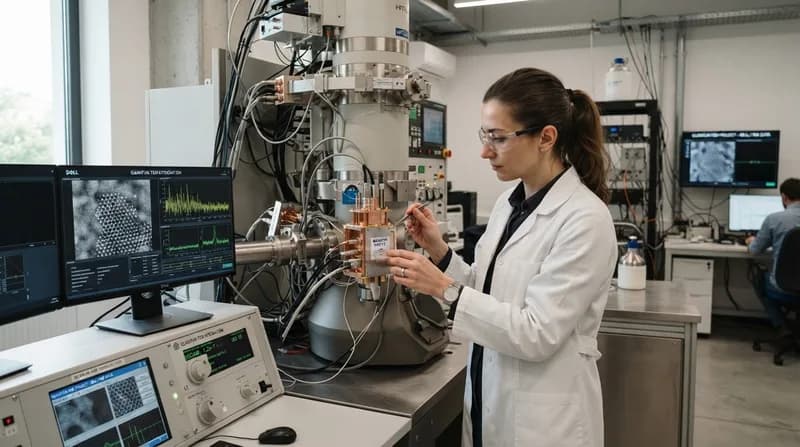
The project is currently transitioning from theoretical modeling to physical construction at TU Wien’s University Service Center for Transmission Electron Microscopy (USTEM). The team, which includes experts from the University of Vienna, JKU Linz, and the University of Innsbruck, has already secured significant support through the 'quantA' Cluster of Excellence and the Gordon and Betty Moore Foundation. The quantum hardware itself, sourced from the University of Innsbruck, is being carefully calibrated to sync with the electron beam path.
If the experimental results mirror the team's mathematical proofs, this hybrid approach could redefine the standards of nanotechnology and structural biology. By allowing researchers to 'read' the quantum information inherent in every electron, this innovation marks a significant leap in our ability to probe the fundamental fabric of reality, proving that the most powerful way to see the smallest things might require the most advanced computers in existence.