A New Era for Connectomics
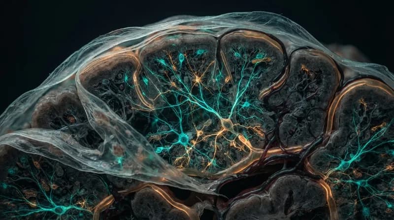
Mapping the human brain is one of the most ambitious challenges in modern science. While neuroscientists have long relied on MRI scans and traditional electron microscopy to chart the brain's massive neural highways, these methods have historically left gaps in our understanding. Now, a collaborative team from Harvard Medical School and the University of California San Diego has achieved a breakthrough: a high-speed imaging approach that maps brain tissue with unprecedented nanoscale detail.
Published in the journal Nature Methods, this study details how the researchers have significantly optimized electron tomography. By refining how data is captured, the team can now image thicker tissue samples using drastically fewer images than conventional methods required. This leap in efficiency allows for a comprehensive 3D view of the brain that was previously computationally and temporally prohibitive, providing a clearer look at the microscopic architecture that governs our thoughts, behaviors, and health.
The Tech Behind the Discovery
The core of this innovation lies in the methodology of electron tomography. Traditionally, this technique involves capturing hundreds of images from various angles to reconstruct a single 3D view, a process that is famously slow and resource-intensive. The research team found a way to achieve a 3D resolution of 15 nanometers or finer using approximately 50 times fewer images than standard protocols.
Furthermore, this new approach is optimized for tissue sections that are roughly 20 times thicker than those typically used in traditional imaging. By reducing the number of required images while simultaneously increasing the volume of the sample, the scientists have made it possible to observe complex cellular interactions that were previously missed by standard mapping techniques.
Why It Matters
- Identifying Electrical Synapses: The imaging revealed previously elusive electrical synapses, or gap junctions, which facilitate near-instantaneous signaling between neurons to synchronize network activity.
- Visualizing Cellular 'Nibbling': The study identified tiny, uncharacterized protrusions between neurons and glial cells, suggesting that neurons may be actively "nibbling" material from their neighboring support cells—a discovery that opens new doors in neurobiology.
- Foundation for Future Research: By creating more accurate connectomes, this technology provides researchers with the tools to investigate neurodevelopmental, cognitive, and neurodegenerative disorders with greater precision.
Future Implications
While the current success is limited to mouse cerebellum tissue, the long-term objective of the team is to scale this technology to map entire mammalian brains. Accomplishing this will require further advancements in automation, particularly in the handling of tissue samples to maintain high-throughput mapping. Once achieved, this technology will offer a transformative look at how the brain matures, how it changes over the course of a human lifespan, and exactly what goes wrong at the cellular level during the onset of neurological diseases. As neuroscientists continue to bridge the gap between structure and function, these high-resolution maps are poised to become the definitive blueprints for the next generation of brain research.