Unlocking the Mystery of Embryonic Development
Congenital heart disease remains one of the most pressing challenges in pediatrics, affecting roughly two out of every 100 newborns globally. While the prevalence of these structural abnormalities is well-documented, the precise molecular triggers have remained elusive. A landmark study from the University of Copenhagen has shed new light on this mystery, revealing that a previously overlooked communication system on the surface of our cells—dubbed a cellular antenna—plays a critical role in how the heart forms during the earliest stages of life.
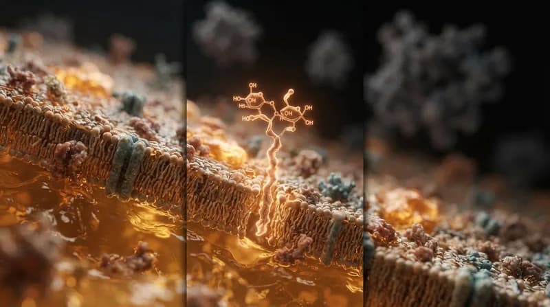
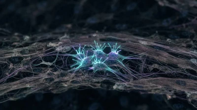
The research team identified a sophisticated signaling hub located within the primary cilium, a microscopic, antenna-like projection found on the surface of most human cells. This structure acts as a sensory organ, interpreting chemical signals from the environment to instruct cells on how to divide, move, or transform into specialized tissues, such as heart muscle. By uncovering this mechanism, scientists are finally beginning to map the 'cog' that, when faulty, leads to severe developmental consequences.
The Role of the Signaling Hub
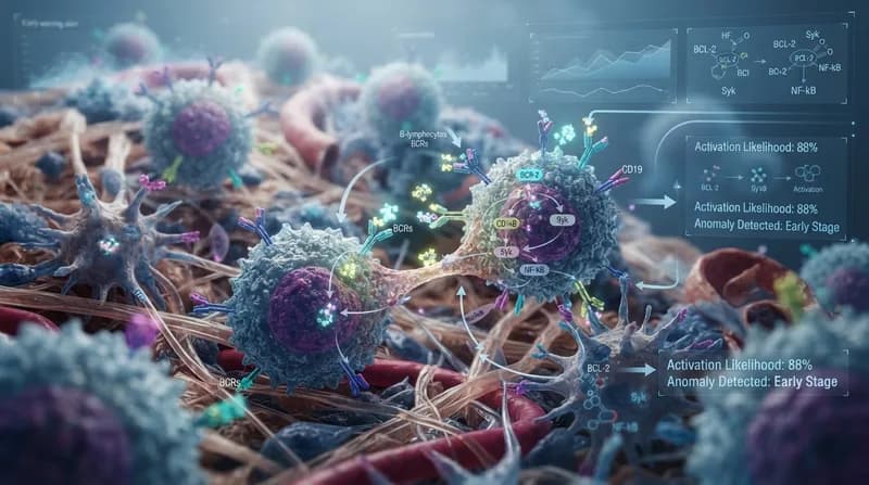
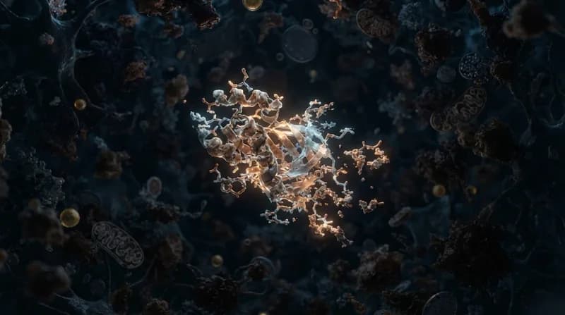
At the center of this discovery is a trio of proteins: TAK1, TAB2, and PKA-Cα. These proteins function collectively within the primary cilium to relay instructions that dictate the fate of embryonic stem cells. Under normal conditions, these molecular signals ensure the seamless development of the cardiovascular system. However, when rare genetic mutations occur, this signaling pathway is disrupted, leading to what researchers call 'antenna defects.'
These genetic malfunctions effectively break the chain of command, preventing stem cells from receiving the necessary cues to develop into functional heart muscle. Because these primary cilia are responsible for sensing critical growth factors and hormones, the resulting disruption doesn't just affect the heart. It provides a biological explanation for why many children with syndromic congenital heart disease also exhibit complications in their brain, kidneys, and skeletal development.
Methodology and Future Implications
To validate this finding, the team utilized a multi-disciplinary approach, cross-referencing genetic data from thousands of patients with congenital heart defects against experimental models. By recreating identified mutations in zebrafish and mouse stem cell models, the researchers observed firsthand how these specific genetic deviations hindered normal heart function and structural formation. This convergence of clinical data and laboratory experimentation offers compelling evidence of the mechanism's role in human development.
Why it Matters
- Unified Diagnosis: This discovery provides a unifying theory for 'syndromic' heart defects, where multiple organ systems are affected simultaneously.
- Early Detection: By identifying the role of primary cilia in these specific genetic profiles, medical professionals may eventually be able to screen for these vulnerabilities earlier in pregnancy.
- Targeted Therapy: Understanding the molecular pathways of the primary cilium opens the door to future genetic-based interventions aimed at correcting or mitigating the impact of these mutations before birth.
While the study, published in PLOS Biology, is a significant leap forward, the researchers emphasize that they are still mapping the full extent of how this mechanism behaves in human physiology. As they continue to explore how these 'tiny antennas' regulate cellular behavior, the findings could ultimately pave the way for a deeper understanding of a wide range of rare genetic conditions that have long baffled the medical community.