The Dual-Origin Revelation
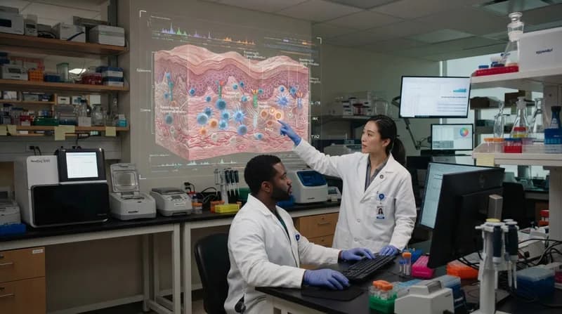
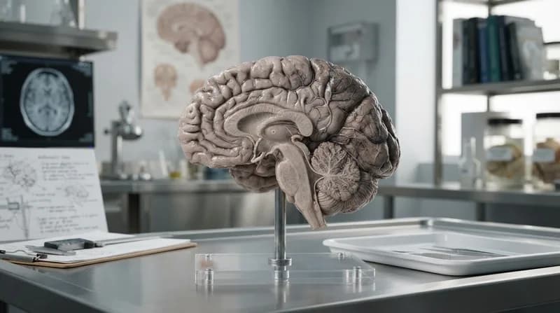
For centuries, the scientific consensus held that the human brain was a unified organ, growing from a singular progenitor cell during early embryonic development. However, groundbreaking research led by Stanford Medicine has effectively dismantled this theory. By examining the earliest stages of embryonic growth, researchers have discovered that the human brain is not a singular entity, but rather a fusion of two distinct, ancient nervous systems that have evolved separately over hundreds of millions of years.
The study, published in Nature Neuroscience, reveals that the front of the brain and the back of the brain originate from entirely different progenitor cell populations. The forebrain and midbrain, responsible for high-level cognitive tasks like consciousness and language, stem from progenitors expressing the Otx2 gene. Meanwhile, the hindbrain, or brain stem—which governs critical autonomic processes like heart rate, breathing, and swallowing—originates from cells expressing the Gbx2 gene. These two cell groups function on parallel, mutually exclusive tracks, effectively acting as separate biological blueprints from the moment of inception.
Solving a Decades-Long Laboratory Hurdle
This fundamental split in brain development explains why scientists have historically struggled to cultivate specific brain cells in a laboratory environment. For years, researchers attempted to coax forebrain-derived stem cells into becoming hindbrain neurons, a task that now appears to be biologically impossible because the two cell types are programmed with distinct chromatin configurations early in development. The inability to replicate hindbrain tissue has been a major barrier to medical progress, particularly for diseases that target the brain stem.
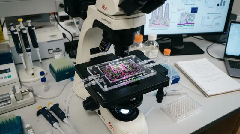
By identifying the specific progenitor cells for the hindbrain, the research team at Stanford has finally succeeded in generating functional human hindbrain motor neurons in a petri dish. These lab-grown neurons exhibit authentic electrical activity and the necessary protein production to regulate vital functions. This technical achievement is transformative, as it provides a live, studyable model of human brain stem tissue for the first time, enabling researchers to observe the progression of diseases that were previously inaccessible for clinical study.
Why It Matters: New Horizons in Medicine
- Targeted Disease Research: The model allows for unprecedented study of Spinal Muscular Atrophy (SMA) and Amyotrophic Lateral Sclerosis (ALS), potentially accelerating the development of regenerative therapies for these conditions.
- Evolutionary Insights: The discovery suggests that, hundreds of millions of years ago, evolution took two separate neural systems—similar to those found in simple organisms like acorn worms—and combined them into the structure we recognize today.
- Obesity and Metabolism: Because the hindbrain contains the circuits responsible for regulating hunger, this breakthrough provides a new platform to study how weight-loss medications interact with the nervous system.
Looking ahead, this dual-organ model shifts the paradigm of neurology. By understanding the brain as two separate pieces fused together rather than a contiguous whole, scientists can now approach neurological medicine with greater precision. Future investigations will likely focus on the spinal cord's developmental origins and how these parallel neural tracks interact to maintain human life, potentially leading to treatments for conditions where the communication between these two "brains" falters.