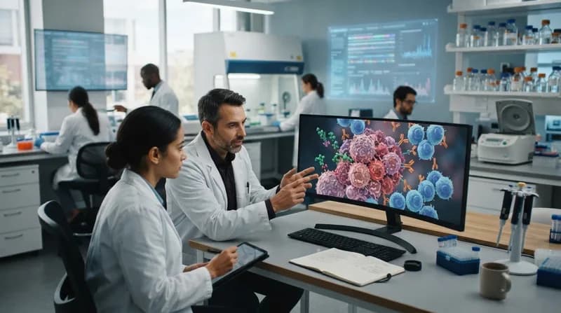
Researchers from the University of Technology Sydney, in collaboration with various partners, have developed a groundbreaking system of smart nanoparticles that significantly improve glioblastoma surgery. These nanoparticles serve a dual purpose: they illuminate hidden cancer cells during surgery and later activate treatments to destroy residual tumor tissue.
The innovative approach was confirmed in mouse studies, where the dual-function system demonstrated a 100% survival rate at 60 days post-treatment. This outperforms traditional methods, highlighting the potential of these smart nanoparticles in combating aggressive brain tumors.
Key Points
- The nanoparticles act as both imaging agents during surgery and a targeted therapy post-operation, activated by near-infrared light.
- Surgeons can visualize tumor clusters as small as 44 micrometers, a substantial improvement over current imaging techniques.
- Post-surgery, the nanoparticles utilize light to convert hydrogen peroxide present in the tumor into oxygen, targeting the low-oxygen environment that often protects cancer cells.
- In glioblastoma studies, all treated mice survived to 60 days, compared to a maximum of 42 days for those undergoing surgery alone.
- This technology is still under early research stages; further studies are essential before human clinical trials can commence.
This revolutionary technology could redefine treatment protocols for glioblastoma by minimizing residual tumor risk and enhancing patient outcomes. However, as emphasized by the researchers, significant steps remain before this methodology can transition from laboratory to clinical application.