A Breakthrough in Micro-Vascular Imaging
Cardiovascular disease remains the world's leading cause of mortality, with atherosclerosis—the accumulation of fatty plaques within arterial walls—serving as the primary culprit. While current intravascular optical coherence tomography (IV-OCT) has significantly advanced how clinicians visualize these blockages, these existing systems are largely restricted to larger arteries. A collaborative team from the Nanjing University of Aeronautics and Astronautics and Nanjing University Medical School has unveiled a groundbreaking piezoelectric microprobe that effectively shrinks the hardware, allowing for unprecedented access to the body’s most delicate vascular networks, including those within the human brain.
Traditional IV-OCT technology relies on near-infrared light delivered via a catheter, requiring 360-degree rotation of the sensor to capture a cross-sectional view of the vessel wall. Current systems utilize either proximal rotation, which suffers from friction-induced distortion in tight spaces, or distal motors, which often create visual artifacts that block the field of view. These limitations have historically constrained catheter diameters to roughly 2 mm, making them unsuitable for the intricate, narrow pathways of the cerebrovasculature.
The Engineering Innovation
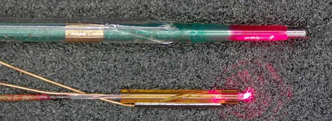
The research team’s new design bypasses these mechanical bottlenecks by utilizing a piezoelectric micro-motor. Measuring just 0.55 mm in diameter, the device is specifically optimized for scanning vessels as small as 2 mm. The mechanism functions by applying a single-phase AC circuit to a piezoelectric crystal, which induces vibrations in a specialized glass tube. A 10-degree groove within this glass then translates longitudinal vibration into torsional, elliptical motion. This ingenious "crank and slider" approach allows the lens to rotate smoothly without the need for bulky electromagnetic motors or complex, wire-heavy drive systems.
Why it Matters: Solving the Rotational Problem
- Reduced Distortion: In laboratory tests against metallic tubes, the probe maintained an angular deviation of just 1 degree at 50 revolutions per second, vastly outperforming traditional proximal catheters that see deviations upwards of 9 degrees.
- Unobstructed View: By moving away from conventional motor-wire layouts, the probe achieves a true 360-degree field of view, ensuring clinicians receive a complete, artifact-free image of arterial health.
- Cerebrovascular Access: The device has already successfully navigated a full-scale model of the human vascular system, reaching the middle cerebral artery, confirming its potential for detecting life-threatening plaques deep within the brain.
- Clinical Precision: When compared against gold-standard histological analyses, the probe’s imaging capabilities accurately identified plaque rupture sites and collagen-rich regions, validating its efficacy as a diagnostic tool.
Clinical Outlook and Implications
The successful integration of this piezoelectric technology marks a significant milestone in diagnostic medicine. By minimizing the probe's footprint and eliminating the mechanical interference that has plagued previous generations of IV-OCT, this technology paves the way for early detection of cerebrovascular diseases that were previously invisible to standard intervention tools. As the team continues to refine this micro-motor architecture, the potential to transition from ex vivo models to human clinical trials suggests that a new standard for high-resolution, minimally invasive vascular diagnostics is on the horizon.