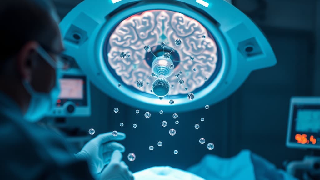
Researchers at the University of Technology Sydney have pioneered a groundbreaking approach to glioblastoma surgery using smart nanoparticles. This innovative system not only illuminates tumor cells during surgery but also delivers postoperative phototherapy to target and eliminate any residual cancer cells. In preclinical trials conducted on mice, this dual-action method resulted in a remarkable 100% survival rate at 60 days.
Key Features of the Nanoparticle System
- Dual Functionality: Operates as both an imaging tool and a therapeutic agent.
- High Precision: Capable of identifying glioblastoma cells as small as 44 micrometers, far exceeding the capabilities of current clinical imaging techniques.
- Safety Profile: In animal trials, there were zero detectable neurological impairments, indicating a high potential for safety in humans.
- Tumor Recurrence: Mice treated with the nanoparticles exhibited no tumor regrowth for up to 60 days, compared to just 42 days observed with standard surgical interventions.
- Mechanism: Utilizes near-infrared light to activate both imaging and therapy, converting tumor-generated hydrogen peroxide into oxygen to alleviate oxygen-poor conditions that often hinder cancer treatments.
This pioneering research holds significant implications for treating glioblastoma, a highly aggressive brain cancer known for poor patient prognosis and limited treatment options. By enhancing surgical precision and improving postoperative outcomes, these smart nanoparticles may pave the way for new therapeutic strategies in oncology.
The findings were originally published in a study titled Smart nanoparticles light up brain cancer and destroy what surgery misses. Further details can be accessed here.