In a significant advancement in the fight against glioblastoma, researchers have created smart nanoparticles that illuminate and destroy cancer cells during and after surgery. This innovative approach, resulting in a 100% survival rate in preclinical studies involving mice, represents a promising avenue for treating this aggressive form of brain cancer.
Key Points
- Glioblastoma has a notoriously low five-year survival rate of only 7%, highlighting the urgent need for better treatments.
- The nanoparticles, developed through collaboration between the University of Technology Sydney, Harvard University, and Henan University, serve dual purposes as both an imaging agent during surgery and a therapeutic treatment afterward.
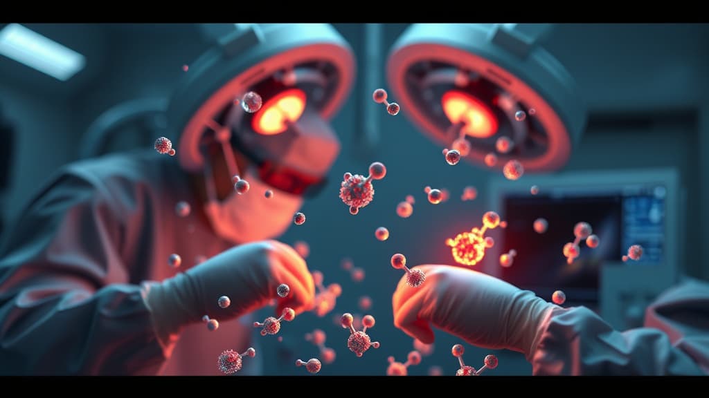
- During operations, surgeons can identify tumor clusters as small as 44 micrometers, thanks to the nanoparticles’ ability to glow under near-infrared light, enhancing the precision of tumor removal.
- In mouse studies, those treated with the nanoparticles survived an average of 60 days post-surgery, a significant increase from 42 days for mice that underwent surgery alone.
- These nanoparticles can penetrate the blood-brain barrier, generating heat and reactive molecules to eliminate residual microscopic cancer cells that surgery might miss.
- While the early results are encouraging, the team emphasizes the necessity for further research to evaluate the effectiveness of this treatment in human patients before progressing to clinical trials.
This dual-function approach not only aids in surgical precision but also ensures that treatment continues even after the operation, potentially changing the landscape of glioblastoma management. The research team remains committed to exploring the nanoparticles' efficacy in human trials, paving the way for future breakthroughs in cancer therapy.