Revolutionizing Digestive Diagnostics
Monitoring the speed at which food travels through the digestive system—a process known as gastrointestinal transit—is a vital tool for diagnosing motility disorders and assessing treatment efficacy. Historically, however, these clinical assessments have relied heavily on imaging techniques like scintigraphy or contrast-enhanced X-rays. While effective, these methods expose patients to ionizing radiation, a significant drawback for frequent check-ups and a major barrier for vulnerable populations such as children and pregnant individuals. A breakthrough collaboration between researchers at IOCB Prague, Charles University, and Ghent University aims to eliminate this risk entirely.
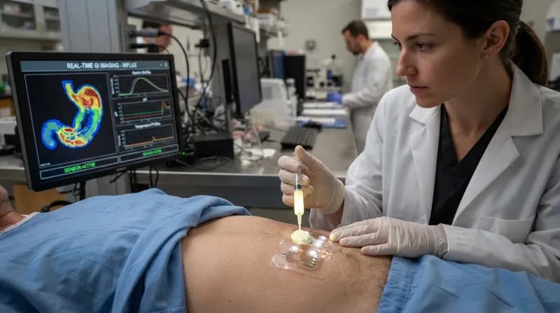
The team has pioneered a novel imaging approach using a water-soluble, fluorinated polymer that acts as a non-invasive contrast agent. By utilizing fluorine-19 magnetic resonance imaging (¹⁹F MRI), clinicians can track this tracer as it navigates the gastrointestinal tract. Because fluorine is virtually absent in the human body, the signal provided by the tracer is exceptionally clean, offering a high-contrast view that lacks the background interference often seen in other imaging modalities.
The Chemistry of Compatibility
One of the most significant hurdles in developing this technology was ensuring the tracer behaved predictably within the stomach and intestines. Previous fluorinated compounds often struggled with hydrophobicity, meaning they would not mix well with the semi-fluid contents of the gut, known as chyme. These materials would often separate into distinct layers, creating imaging artifacts that obscured results rather than clarifying them.
The new, highly hydrophilic polymer overcomes this by blending seamlessly with digestive contents. Finding this balance required precise molecular engineering. The research team focused on maximizing the fluorine concentration to ensure a robust signal for ¹⁹F MRI without compromising the material’s solubility. The result is a stable, reliable tracer that tracks the actual flow of digestive material through the system, providing a real-time, high-definition map of transit times without any associated radiation exposure.
Why It Matters
- Patient Safety: By removing ionizing radiation, the method allows for frequent, longitudinal monitoring of digestive health, opening doors for long-term patient care.
- Inclusivity: The non-radiological nature of the scan makes it suitable for patients previously contraindicated for traditional X-ray-based diagnostic tests, including pregnant women and pediatric patients.
- Diagnostic Precision: Combining anatomical MRI with ¹⁹F MRI overlays provides a clearer picture of both the physical structure of the GI tract and the functional speed of digestion.
From Preclinical Success to Clinical Future
The current proof-of-concept study has yielded promising results in animal models. During the testing phase, the polymer tracer was administered to rats, allowing researchers to successfully monitor its entire journey through the digestive tract until excretion. Histological examinations confirmed that the tracer moved through the system without causing tissue damage or entering the systemic circulation, marking a significant milestone in safety verification.
While the transition from preclinical research to human clinical practice involves a rigorous multi-year path, the implications are profound. The team is already iterating on a next-generation version of the polymer, seeking to secure patents as they prepare for further development. If successful, this innovation could redefine the standard of care for gastroenterology, replacing outdated, radiation-heavy procedures with a safer, high-resolution diagnostic standard that is as precise as it is patient-friendly.