A New Frontier in Cellular Imaging
Researchers at Stanford University have reached a significant milestone in microscopy, unveiling a new system capable of observing the internal machinery of living cells in real-time. The breakthrough technology, dubbed Interferometric Image Scanning Microscopy (iISM), allows scientists to capture detailed footage of cellular structures as they move and interact, all without the need for traditional fluorescent labels.
For decades, biological imaging has relied on chemical markers to highlight specific proteins or organelles. While powerful, these methods come with significant drawbacks, including potential interference with natural cellular behavior, the risk of photobleaching, and the inability to label more than a few structures at a time. The iISM system circumvents these challenges, offering a 'label-free' perspective that provides a more accurate, natural look at the microscopic processes driving life.
The Technology Behind the Lens
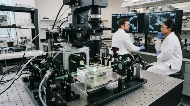
The iISM system is built upon a clever synthesis of two distinct optical technologies. It utilizes interferometric scattering—a method that detects light scattered by tiny structures within a cell—and pairs it with an advanced detector array similar to those found in high-end confocal microscopes. Because scattered light signals are inherently weak, the system employs a secondary laser beam to amplify these signals through interference, resulting in an exceptionally high-contrast image.
By swapping the traditional pinhole and single-detector setup for a multi-view detector array, the microscope gathers hundreds of perspectives of a single region. Advanced computational algorithms then stitch these views together, producing images with a lateral resolution of approximately 120 nanometers. Crucially, the system operates with significantly lower illumination power than other high-resolution methods, ensuring that cells remain healthy and undisturbed during extended observation periods.
Why It Matters
- Non-Invasive Observation: By eliminating fluorescent labels, researchers can observe cells in their most natural, undisturbed states.
- Extended Study Durations: The low-power illumination minimizes photodamage, allowing for longer, continuous imaging sessions.
- Broad Research Applications: The technique provides a powerful, versatile tool for fields ranging from pharmacology to plant biology.
- Complementary Utility: Rather than replacing existing tools, iISM is designed to work alongside fluorescence microscopy to provide both molecular specificity and spatial context.
Real-World Implications for Science and Medicine
The implications for this technology are vast, spanning several critical areas of medical research. Current collaborations at Stanford are already testing the microscope's utility in real-world scenarios. One project involves observing how cancer drugs are processed as they enter a cell, offering a potential window into how medications interact with their targets in real-time. Another study focuses on the physical changes in red blood cells during malaria infections, providing a clearer look at disease progression at the microscopic level.
Furthermore, the technology is being used to explore complex environmental interactions, such as the relationships between plant cells, fungi, and soil microbes. As the researchers continue to refine the instrument, they aim to make the technology accessible to the broader scientific community, potentially opening new pathways for drug discovery and a deeper understanding of human disease. By merging physical precision with computational power, the team has delivered a tool that promises to change how we visualize the very building blocks of life.