The Paradigm Shift in PET Imaging
For decades, PET/CT scans have been a cornerstone of oncological diagnosis and monitoring, but they have long suffered from a significant technical limitation: the short axial field of view. Conventional systems force clinicians to image the body in segmented 'bed positions,' a process that is time-consuming and often uncomfortable for patients. However, recent findings presented at the 39th Annual Congress of the European Association of Nuclear Medicine suggest that this standard is about to be eclipsed by next-generation total-body PET/CT technology.
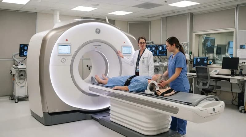
Researchers at the Peter MacCallum Cancer Centre have successfully evaluated a new 128 cm long-axial field-of-view (LA-FOV) system, specifically the GE HealthCare Omni 128 cm. By capturing a vast portion of the human body in a single pass, this hardware detects a significantly higher volume of signals emitted by radiotracers, enabling clinicians to drastically slash exposure times without sacrificing the clinical utility of the resulting images.
The EQUIP Study and Diagnostic Performance
The EQUIP study focused on comparing the performance of this advanced 128 cm system against conventional short-axial field-of-view (SA-FOV) scanners. Over the course of the research, twenty-four participants were scanned using five common radiotracers, including 18F-FDG and 68Ga-PSMA. The study revealed that even when the capture time for the LA-FOV system was compressed to a mere one minute, the diagnostic quality consistently outperformed the standard-of-care scans used in clinical practice today.
In subjective evaluations, nuclear medicine specialists blind-tested the images based on sharpness, lesion conspicuity, and image noise. The results were compelling: the 10-minute and 5-minute scans earned perfect diagnostic scores, while the 1-minute images—despite having slightly more noise—still secured an average score of 4.5 out of 5. This result is particularly striking when measured against the conventional scanner's score of 3.9, proving that speed does not have to come at the expense of precision.
Why it Matters: Implications for Clinical Workflow
- Enhanced Patient Comfort: Shorter scan times are a massive relief for patients suffering from chronic pain or those who struggle to remain perfectly still for extended durations.
- Reduced Movement Artifacts: With scan times dropping from ten minutes to one, the likelihood of image blurring caused by involuntary patient movement or breathing is significantly mitigated.
- Increased Throughput: Imaging departments could potentially process a higher volume of patients per day, reducing wait times for critical cancer diagnostics.
- Flexible Radiotracer Dosing: The superior sensitivity of the LA-FOV system provides clinicians the option to either scan faster or, alternatively, reduce the administered radiation dose to the patient.
As the medical community looks toward the future, these findings indicate that total-body PET imaging is poised to become the new benchmark for efficiency in oncology. While larger, follow-up studies are necessary to ensure that these lightning-fast scans do not overlook faint lesions, the current data suggests that the hardware is already more than capable of surpassing the quality standards that have governed the field for years.