Revolutionizing Precision Surgery
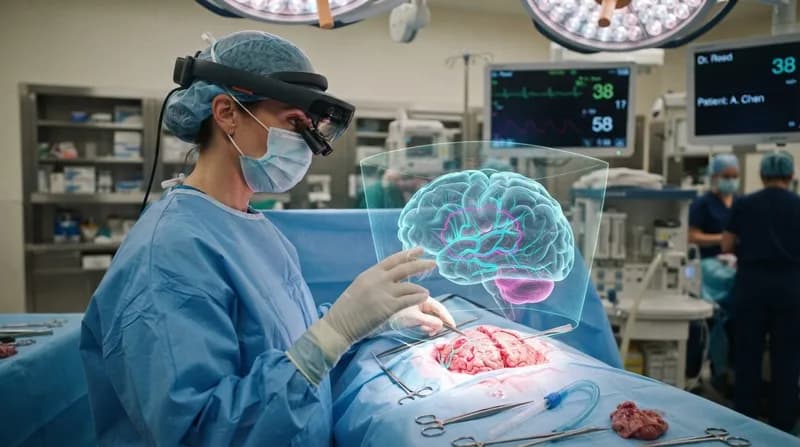
Laparoscopic—or keyhole—surgeries have long been the gold standard for minimally invasive procedures, but they come with a significant challenge: the inability to see beneath the surface of soft organs. Surgeons often rely on pre-operative scans, which are static and do not account for the shifting, soft nature of biological tissues. A new, innovative 3D augmented reality (AR) technology developed at Chalmers University of Technology is now addressing this gap, effectively providing surgeons with a real-time 'internal GPS' during live operations.
By overlaying a dynamic 3D visualization of tumors and underlying anatomical structures directly onto the surgeon's camera feed, the system allows for unparalleled accuracy. As the laparoscopic camera moves, the digital overlay adjusts instantly, ensuring that the surgeon's view remains synchronized with the physical organ. This continuous guidance ensures that hidden tumors, which were previously invisible to the naked eye during standard camera-assisted procedures, can now be precisely targeted and excised with significantly improved confidence.
The Anatomy of the Tech: QR-Powered Navigation
The system relies on a remarkably simple yet ingenious interface: a small, one-square-centimeter sticker featuring a QR code. This marker is placed directly on the surface of the organ—in the case of the initial successful surgery, the liver—acting as an artificial reference point. Because organs like the liver lack distinct surface landmarks and can be as smooth as a polished floor, this marker provides the necessary geometric anchor for the software.
Once the marker is in the camera's field of view, the system fuses the real-time visual feed with pre-operative X-ray and CT data. The software calculates the precise position of the tumor relative to the marker, translating complex mathematical modeling into a clear, augmented visual overlay. During a clinical milestone in June 2026, this system proved to be both stable and intuitive, allowing for a successful tumor resection that permitted the patient to be discharged just one day later.
Why It Matters
- Enhanced Accuracy: Provides real-time, dynamic guidance that static pre-op scans cannot offer.
- Broader Applications: While currently tested on the liver, the underlying math and AR framework are being adapted for various laparoscopic procedures.
- Interdisciplinary Success: The project demonstrates the power of long-term collaboration between medical specialists and applied mathematicians.
- Future Scalability: Plans are already in motion for international studies involving hospitals in Oslo and Washington to validate the system's global utility.
The Future of Interdisciplinary Medical Innovation
The genesis of this technology dates back a decade, emerging from a unique partnership between surgeons at Sahlgrenska University Hospital and mathematicians at Chalmers University of Technology and the University of Gothenburg. The project serves as a masterclass in interdisciplinary collaboration, where engineers and medical professionals worked in tandem to solve a high-stakes, real-world clinical problem. By focusing on the functional needs of the operating room rather than rigid departmental boundaries, the team successfully developed a system that bridges the gap between raw data and actionable surgical insight.
Looking ahead, the development team is working alongside Chalmers Next Labs to broaden the system's reach. As the project moves through clinical validation and regulatory approval processes, the team aims to standardize the technology for a variety of routine surgical procedures. By turning invisible internal threats into clearly defined targets, this AR tool is poised to become a staple in modern operating rooms, potentially reducing complications and improving outcomes for patients worldwide.