The Shift Toward Personalized Vascular Care
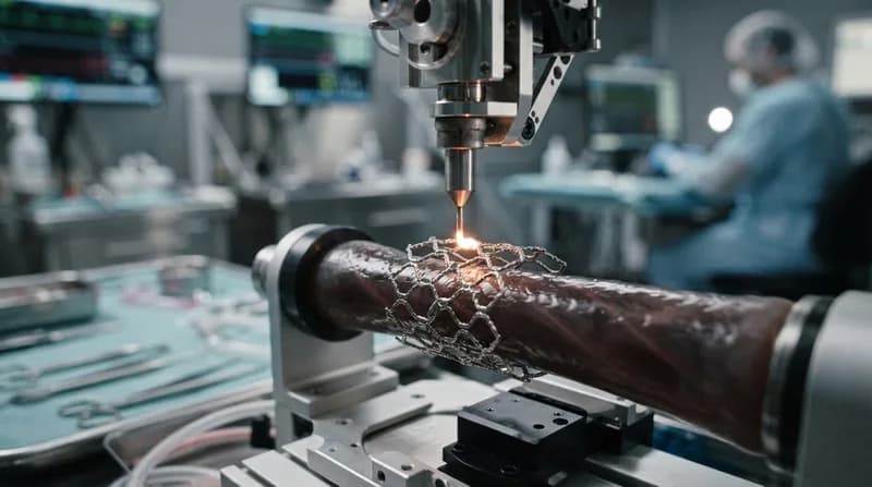
An international team of researchers, led by the IMDEA Materials Institute and the Universidad Politécnica de Madrid, has unveiled a pioneering method for the additive manufacturing of cardiovascular devices. By 3D-printing nitinol—a highly flexible, superelastic shape-memory alloy—the team can now produce complex, personalized implants that adapt perfectly to a patient’s specific anatomy. This breakthrough, recently published in npj Metamaterials, offers a solution to the long-standing physical limitations of traditional medical device manufacturing.
The technology addresses a critical bottleneck in the production of minimally invasive transcatheter devices (MITDs). Historically, devices like stents were restricted to rigid, repetitive lattice structures. While functional, these designs often struggle to manage the extreme physical stress of being compressed into a catheter, navigated through winding blood vessels, and then deployed at the target site. This new methodology overcomes those constraints by enabling the creation of intricate, braided geometries in a single, continuous production step.
Overcoming Structural Limitations with Braided Metamaterials
Standard additive manufacturing typically utilizes lattice structures composed of rigid, interconnected struts. During clinical procedures, these structures often fail or become too stiff when subjected to high-strain environments, increasing the risk of vessel injury or device fracture. The research team’s new approach abandons these rigid struts in favor of braided, interwoven filaments that behave more like textile meshes than traditional metal framework.
By utilizing a sophisticated post-processing technique involving chemical etching and electropolishing, the researchers reduced the fiber thickness of their printed designs by nearly 48%. This refinement allowed the stents to achieve remarkable performance metrics. During testing, the braided prototypes successfully endured compressions of up to 50% of their diameter, followed by a 90% elastic recovery rate. Perhaps most significantly, the chronic outward force exerted by these devices on arterial walls was reduced by up to five times compared to conventional designs, greatly minimizing the risk of post-implantation thrombosis or vascular damage.
Why It Matters
- Patient-Specific Design: Implants can be reconstructed directly from patient CT scans to ensure an exact fit.
- Extreme Flexibility: Braided nitinol structures can navigate complex, tortuous vascular pathways without fracturing.
- Lower Clinical Risks: Reduced chronic outward force minimizes vessel trauma and inflammation.
- Material Innovation: The process represents a major evolution in how superelastic alloys are utilized in life-saving medical tech.
Future Directions in Neurovascular Medicine
While the initial prototypes have proven successful in controlled mechanical testing, the path toward clinical application involves several more phases, including in-vitro biocompatibility assessments and animal studies. However, the potential for this technology extends far beyond simple stent applications. The research team is already setting its sights on the next frontier: the human brain.
Developing medical devices for neurovascular intervention requires extreme precision to navigate the delicate and highly restricted regions of the brain. The team plans to explore ultra-high-precision printing methods, such as two-photon polymerization, to miniaturize these complex nitinol structures further. Additionally, the integration of magnetic functionalization could eventually allow for remote, catheter-free navigation of these devices, marking a new era in minimally invasive neurosurgery. By successfully winning the National James Dyson Award, this research has already signaled its immense promise to the global medical community.