Decoding the Early Neural Response to Antidepressants
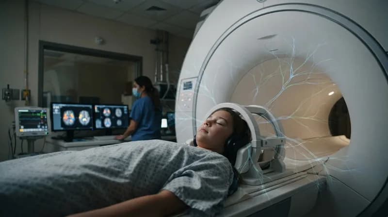
Major depressive disorder (MDD) has long challenged clinicians, largely because the efficacy of antidepressant medications is often unpredictable and slow to manifest. A recent study published in Nature Mental Health aims to bridge this diagnostic gap by tracking how the brain responds to treatment within the first two weeks. By analyzing resting-state functional magnetic resonance imaging (fMRI) scans of 386 participants, researchers from Stanford University, the University of Texas at Austin, and other institutions have identified distinct patterns of brain connectivity that emerge shortly after treatment begins.
The study specifically examined patients receiving either a placebo, sertraline, or escitalopram—two of the most common selective serotonin reuptake inhibitors (SSRIs). By comparing baseline scans with those taken one to two weeks into the treatment cycle, the research team employed advanced machine learning frameworks to isolate changes in functional connectivity (FC), defined as the synchronization of activity between different brain regions.
The Universal Signature: Visual and Thalamic Connectivity
One of the most surprising findings was a set of brain changes that occurred regardless of the specific treatment administered. Across all groups—including those who received the placebo—the researchers observed increased connectivity within a neural system involving the visual cortex, the precuneus, and the thalamus. The thalamus, a critical relay center deep within the brain, appeared to sync more closely with regions responsible for visual processing and the precuneus, an area associated with self-referential thought and memory.
This generalized response suggests that the mere act of entering a clinical treatment program may trigger a common neural shift, independent of the pharmacological intervention. While these changes were not strictly tied to clinical improvement, their presence across the board highlights a foundational brain mechanism that activates early in the therapeutic process.
Targeted Changes: The Drug-Specific Effects
Beyond the universal neural shifts, the researchers identified specific biomarkers unique to the SSRIs. These changes were found in a subset of patients who received sertraline or escitalopram, localized in regions critical to emotional regulation and executive function, including the amygdala, the midcingulate cortex, the orbitofrontal cortex, and the cerebellum. This subset-specific activity provides a mechanistic look at how antidepressants interact with the brain’s emotional processing centers.
Interestingly, the study noted that patients who did not show these drug-specific changes could be effectively identified using predictive models built on their placebo response. This implies that the brain’s initial reaction to a placebo versus a drug can act as a barometer for how a patient might eventually respond to medication.
Why It Matters
- Personalized Medicine: Identifying early biomarkers allows clinicians to predict whether a patient will respond to a specific SSRI within two weeks, potentially saving months of trial-and-error.
- Neuro-Mechanistic Insight: The differentiation between placebo-mediated changes (often tied to the striatum and attention networks) and drug-specific changes helps clarify the physical roots of antidepressant efficacy.
- Predictive Modeling: The use of machine learning in this study sets a precedent for using neuroimaging as a diagnostic tool in psychiatry, moving beyond subjective symptom reporting.
While the study represents a significant step forward in understanding the neural architecture of depression, the researchers emphasize that further validation is necessary before these scans can become a standard clinical tool. Nevertheless, the ability to observe such precise, treatment-induced changes at the two-week mark offers a promising new frontier in the quest to optimize mental health treatment.