Bridging Biophysics and Oncology
A collaborative research effort between the Institute of Science and Technology Austria (ISTA) and the Francis Crick Institute in the UK has reached a critical breakthrough in understanding cancer metastasis. By applying advanced microfluidic technology, the team has successfully modeled the complex microenvironments that surround tumours, revealing how physical disorder in the body can act as a catalyst for cancer cells to break away from their collective and invade healthy tissue.
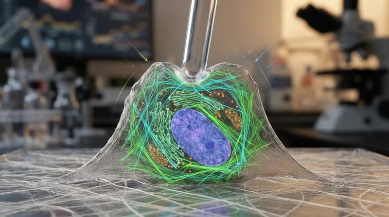
Metastasis remains the primary driver of mortality in cancer patients. While genetic mutations play a significant role, the physical environment surrounding a tumour—including connective tissue, blood vessels, and immune cells—is equally pivotal. The research team sought to replicate the difficult physical conditions cancer cells encounter as they migrate through the body by designing experiments that mimic the dense, fibrous landscapes typical of human tissue.
The Microfluidic Chip Design
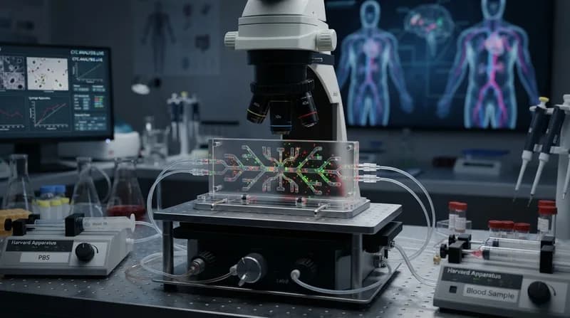
To investigate these dynamics, researchers utilized specialized microfluidic chips fabricated on silicon wafers. Leveraging standard semiconductor lithography, the team engineered "forests" of microscopic pillars. These structures act as artificial obstacles that tumour cells must navigate. By creating both perfectly square, ordered lattices and intentionally disordered patterns—where pillars were shifted by random angles and distances—the researchers were able to quantify how geometry influences cellular behavior.
Saren Tasciyan, a biotech data science consultant who developed the architecture, had to write bespoke software to define these mathematically disordered landscapes. This allowed for a precise, reproducible study of how heterogeneity affects cell detachment. The findings were clear: in environments with high levels of disorder, tumour cell collectives were significantly more prone to breaking apart, a process that directly mirrors the initial stages of metastasis.
Computational Modeling and Future Implications
To expand on these experimental observations, the team paired their physical chip data with sophisticated computer simulations. Postdoc Zuzana Dunajová modeled individual cancer cells as interacting beads moving through varying geometric environments. These simulations confirmed that the effects observed on the microfluidic chips are cumulative, with the irregular surfaces of the environment causing the cell interface to develop finger-like protrusions. These protrusions serve as the primary exit points for individual cells to detach from the main tumour mass.
- Enhanced Understanding: The study highlights that the physical "roughness" or disorder of a tumour's surroundings can be a direct driver of malignancy.
- Technological Synergy: By combining semiconductor manufacturing with biological research, the team has created a scalable, precise model for studying cell dynamics.
- Strategic Insight: The research suggests that physical, rather than just genetic, factors are fundamental to how a cancer becomes invasive, opening new doors for therapeutic targeting.
This study provides a robust biophysical model that not only clarifies how cancers spread but also emphasizes the importance of the microenvironment in cancer progression. By standardizing the measurement of environmental disorder, this research provides a new toolkit for oncologists and biologists to study the mechanics of cancer movement in a controlled, high-precision environment.