The Evolving Role of PET Imaging in Neurology
Nuclear medicine is currently undergoing a paradigm shift in the fight against Alzheimer’s disease. As new monoclonal antibody therapies like lecanemab and donanemab gain traction, the ability to accurately identify candidates for treatment has become a critical clinical challenge. Experts at the 39th Annual Congress of the European Association of Nuclear Medicine are emphasizing that amyloid and tau PET imaging are no longer just research tools; they are now vital instruments for routine clinical decision-making, enabling a new era of precision neurology.
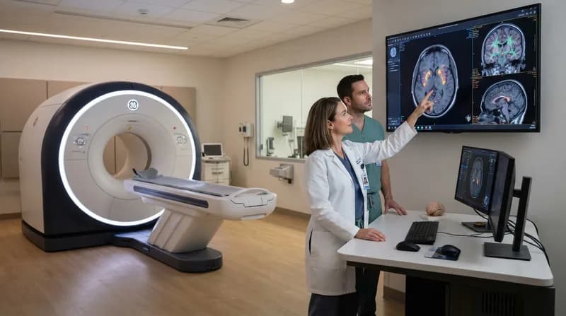
By utilizing targeted radiotracers, clinicians can visualize biological changes in the brain that are invisible to standard imaging modalities. This granular view allows medical professionals to confirm amyloid pathology—the hallmark of Alzheimer’s—and distinguish it from other forms of dementia. This diagnostic precision is essential, as the latest generation of anti-amyloid treatments is only effective when targeting specific beta-amyloid aggregates associated with Alzheimer’s pathology.
Amyloid PET: Diagnosis and Treatment Monitoring
Amyloid PET scans serve a dual purpose: they act as a gatekeeper for starting therapy and a metric for determining when to potentially halt it. Because amyloid plaque buildup begins years before cognitive symptoms emerge, early detection via PET imaging is critical for capturing the window of opportunity where therapeutic interventions can slow disease progression by up to 30%. By confirming the presence of plaques, doctors can ensure that high-cost, intensive monoclonal antibody treatments are directed at the patients most likely to see meaningful clinical benefit.
Furthermore, these scans offer a quantitative way to track treatment efficacy. Clinical data suggests that many patients reach an 'amyloid-negative' status after roughly 12 months of donanemab therapy. In these instances, PET imaging provides the clinical justification to consider ending a drug regimen, potentially sparing patients from unnecessary long-term exposure and health system resources. As trials continue to refine these protocols, PET imaging is emerging as the gold standard for personalized treatment duration.
Tau PET and the Future of Predictive Care
While amyloid PET confirms the disease, tau PET imaging provides the roadmap for how the disease will behave. Tau protein accumulation is closely linked to the disruption of nerve cell communication, and its presence serves as a primary marker for neuronal damage. Clinicians now use tau PET to assess the extent of existing brain damage, which helps predict how a patient will respond to amyloid-targeting drugs. Current data indicates that patients with lower tau levels typically show a more robust response to modern monoclonal antibodies, making tau scans indispensable for managing expectations and selecting therapy candidates.
Why It Matters
- Precision Medicine: Eliminates guesswork by matching drugs to the specific biological state of the patient.
- Resource Efficiency: Enables clinicians to stop treatments once amyloid clearance is achieved, reducing unnecessary costs.
- Early Intervention: Provides a window into the brain years before significant cognitive decline begins.
- Prognostic Power: Tau imaging provides clearer insights into disease progression, helping families plan for the future.
The Next Frontier: Trontinemab
Looking ahead, the development of new antibodies is promising even faster intervention. A prime example is trontinemab, currently in phase 3 trials, which utilizes a specialized 'shuttle' mechanism to cross the blood-brain barrier more effectively. If approved, this could potentially shorten the intensive phase of treatment from over a year to just three or four months. In this future landscape, PET imaging will remain the essential tool for validating treatment success, ensuring that medical progress is guided by concrete evidence of physiological improvement.