A New Frontier in Respiratory Research
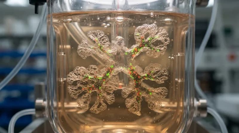
In a significant leap for medical science, researchers at the Stanford University School of Medicine have successfully developed human lung organoids that feature an integrated, functional immune system. Published in the journal Nature, this breakthrough allows scientists to study the body's primary defenses against pathogens in a controlled, 3D environment that mimics the complex reality of human lung tissue. By utilizing distal lung tissue from over 220 patient donors, the team created a sophisticated air-liquid interface culture that supports the long-term survival of critical immune cells.
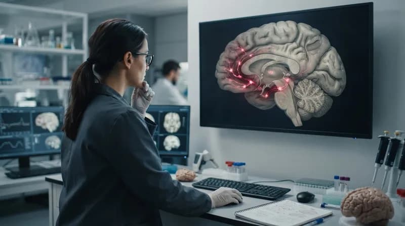
The hallmark of this advancement is the presence of resident T cells that remain within the tissue, maintaining their ability to detect and respond to viral threats. Unlike traditional cell cultures that lack the depth of a living organism, these organoids enable investigators to isolate and examine local tissue immunity without the confounding variables introduced by the rest of the body’s systemic immune responses. This high level of control provides a promising alternative to animal testing, potentially offering insights into human biological responses that were previously difficult, if not impossible, to capture in traditional laboratory settings.
Validating Immune Function and Adaptive Memory
To confirm that these organoids were more than just passive structures, the research team exposed them to SARS-CoV-2. The results were telling: the organoids mounted both generalized inflammatory responses and highly specific, adaptive immune reactions. The resident T cells demonstrated a clear ability to recognize and target the virus, proving that the immune components within the model were not only present but fully operational. This responsiveness allows for the precise study of how our lungs identify and neutralize pathogens on the front lines.
Furthermore, the researchers explored the potential for "local vaccination" strategies. By pre-stimulating the organoids with viral fragments, the team effectively trained the tissue's immune cells to identify the virus before a full-blown infection occurred. This stimulation resulted in significantly weakened subsequent infections, providing empirical evidence that training the lung’s local defenses is a potent strategy for disease mitigation. This discovery suggests that future therapies could focus on vaccines delivered directly to the respiratory mucosa, potentially acting as a powerful complement or alternative to systemic immunization.
Why It Matters
- Reduces reliance on animal models: These organoids offer a more accurate human-centric platform for drug and vaccine testing, potentially increasing the success rate of clinical trials.
- Deepens understanding of tissue-resident immunity: The study reveals that T cells in the lungs are more persistent than previously assumed, shifting our understanding of long-term respiratory defense.
- Broad therapeutic application: While the study utilized SARS-CoV-2 as a test case, the platform is adaptable for investigating complex conditions like asthma, COPD, and bacterial infections, paving the way for personalized medicine.
Looking ahead, the development of these immune-integrated organoids promises to accelerate the pace of respiratory research. By isolating the mechanics of local lung immunity, the scientific community now has a robust, longitudinal model to test everything from novel antiviral agents to personalized pulmonary treatments. This innovation marks a fundamental shift toward more efficient, accurate, and ethical methodologies in the study of global health challenges.