Revolutionizing Cardiology with Non-Invasive Diagnostics
A team of researchers from the Universitat Politècnica de València (UPV), in collaboration with international partners, has unveiled a sophisticated artificial intelligence framework that could fundamentally change how we diagnose and manage atrial cardiomyopathy. By utilizing graph neural networks (GNNs), the team has demonstrated the ability to pinpoint and quantify structural abnormalities in heart tissue using nothing more than electrical signals collected from the surface of a patient's torso.
Atrial cardiomyopathy, characterized by electrical and structural changes like fibrosis, is a primary driver of atrial fibrillation—one of the world's most prevalent heart rhythm disorders. Historically, identifying the specific location and extent of this tissue damage has required invasive procedures, such as intracardiac electroanatomical mapping, or expensive and time-consuming MRI scans. This new AI-driven approach offers a potentially game-changing, non-invasive alternative for clinical assessment.
How the Technology Functions
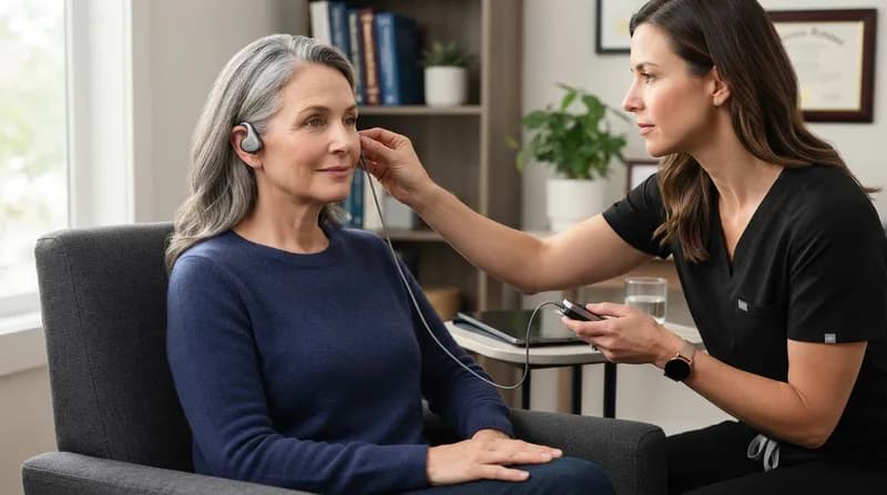
The core of this advancement lies in the processing of Body Surface Potential Maps (BSPMs). By placing electrodes across the torso, clinicians can record the heart's complex electrical activity. The researchers trained their GNN model on a massive dataset of 14,400 simulated electrical maps, which encompassed a wide range of diverse atrial and torso anatomies, as well as varying degrees of pathological tissue damage.
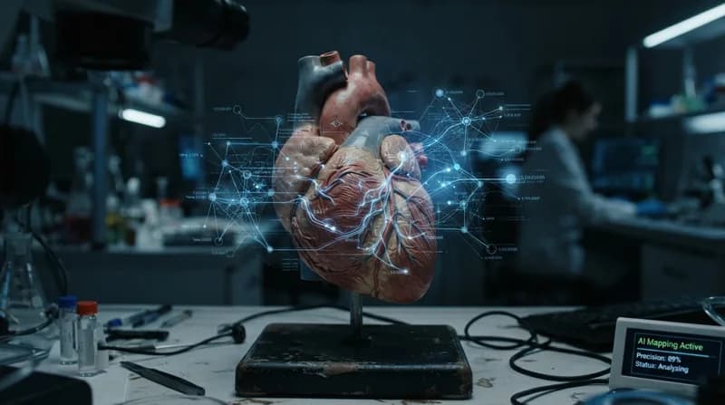
Unlike traditional linear analysis, the graph neural network excels at interpreting the spatial distribution and temporal evolution of electrical signals across the body. The model essentially "maps" the surface potential data back to the underlying anatomy of the heart. The results are highly encouraging: in initial testing, the system achieved 89% accuracy in localizing affected atrial tissue, accompanied by 89% sensitivity and 90% specificity. Furthermore, the model was able to determine the extent of tissue damage with 84% accuracy, maintaining stable performance even when subjected to signal noise.
Why It Matters
- Non-Invasive Potential: Reduces the need for risky, invasive cardiac catheterization procedures.
- Robustness: The model demonstrated an impressive ability to generalize its findings to anatomies it had never encountered during the training phase, which is critical for real-world patient diversity.
- Clinical Planning: Precision mapping of damaged tissue could allow cardiologists to better plan interventions, such as ablation procedures, by identifying the exact "hotspots" that require treatment.
The Path to Clinical Implementation
While the results published in the journal Discover Computing are highly promising, the research team emphasizes that this is currently a proof-of-concept study. The model has been refined using high-quality simulated data, which provides a clean baseline for training. However, the transition from a laboratory simulation to a hospital setting requires rigorous validation using real-world patient clinical records.
Looking ahead, the team is focused on prospective validation trials. If the model proves as effective with human patient data as it has with simulations, it could become a standard tool in cardiology suites worldwide. By providing a clear, accurate, and non-invasive window into the health of atrial tissue, this AI model represents a significant leap forward in the ongoing quest to treat cardiac arrhythmias with greater precision and patient comfort.