A New Frontier in Neuro-Cartography
In a monumental achievement for neuroscience, researchers at the University of California, Irvine, have successfully mapped approximately 8 million individual cells within the human basal ganglia. By identifying 60 distinct cell types and documenting their specific spatial communities, this study offers the most granular look yet at the deep-brain regions responsible for governing movement, motivation, and decision-making. This work, published in the journal Cell, serves as a cornerstone for future research into neurodegenerative conditions like Parkinson’s and Huntington’s disease.
Led by the Center for Neural Circuit Mapping, the initiative marks a departure from traditional single-cell sequencing. While standard sequencing excels at identifying cell types, it often strips away the vital context of a cell’s physical neighborhood. The UC Irvine team bridged this gap by employing spatial transcriptomics—specifically utilizing MERFISH+ and Stereo-seq technologies—to pinpoint exactly where these cells reside within the basal ganglia’s intricate anatomy. By retaining this spatial information, scientists can now visualize how specific "cellular communities" form and function in the healthy human brain.
The Anatomy of Cellular Communities
The study reveals that the organization of the basal ganglia is far more structured than previously understood. Researchers discovered that specific cell types occupy defined compartments, with some supporting cells clustering exclusively along regional borders. Perhaps most intriguingly, the team identified particular neuron subtypes that appear uniquely abundant in primates, localized within specific compartments of the basal ganglia. This spatial organization suggests that brain function is not just about the types of cells present, but the specific configurations and interactions they maintain within their environment.
Understanding these "cellular neighborhoods" provides a critical baseline for comparative studies. By having a high-resolution reference of what a healthy brain looks like, researchers are now positioned to investigate why certain cell populations are susceptible to disease while others show resilience. This level of detail helps scientists determine if a cell's neighbors play a protective or destructive role, potentially uncovering new therapeutic targets for complex neurological and psychiatric disorders.
Building a Sustainable Research Infrastructure
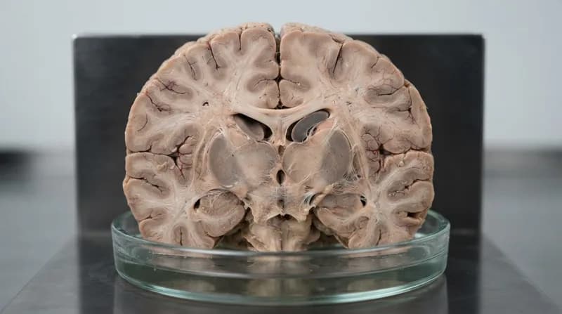
Beyond the map itself, the researchers addressed the foundational logistical hurdles of human brain research. A companion study published in Cell Systems details the complex pipeline required to procure, prepare, and characterize high-quality human brain tissue. This infrastructure is essential for maintaining the rigorous standards necessary for molecular research. Through collaborations with forensic and medical institutions, the team has established a standardized workflow that ensures anatomical documentation and tissue integrity, which are vital for the validity of large-scale mapping projects.
Why It Matters: Context for the Future
- Foundation for Discovery: Much like the Human Genome Project, this atlas provides a shared reference point for the global scientific community.
- Disease Vulnerability: The ability to compare healthy tissue against diseased tissue allows researchers to isolate specific cellular failures, offering a roadmap for precision medicine.
- Standardized Methods: By establishing unified classification methods, the project allows findings from diverse labs and species to be compared accurately, accelerating cross-disciplinary discovery.
- Multi-Institutional Collaboration: Part of the wider BRAIN Initiative Cell Atlas Network (BICAN), this effort integrates expertise from 55 institutions worldwide, ensuring that the methodologies developed here will guide brain mapping efforts for decades to come.