The Mechanics of Ultrasonic Tissue Breakdown
Histotripsy is a non-invasive medical technology that leverages focused ultrasound to mechanically break down targeted tissue using cavitation. Unlike traditional thermal ablation, which relies on generating heat to destroy cells, histotripsy utilizes the rapid expansion and collapse of micro-bubbles to create intense mechanical stress. While this process is highly effective for many soft tissue types, it has historically faced challenges when dealing with fibrous, mechanically resistant tissues, such as those found in benign prostatic hyperplasia (BPH). These tissues, dense with smooth muscle and collagen, often resist standard treatment protocols, prompting researchers to investigate whether the timing of ultrasound delivery is as critical as the intensity.
The Duty Cycle Fallacy
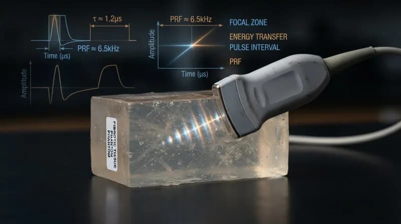
In clinical practice, ultrasound exposure is frequently summarized by the 'duty cycle'—the fraction of time the device is actively emitting energy. A recent study published in Scientific Reports challenges the assumption that treatments with the same duty cycle will yield identical results. By maintaining a constant 1% duty cycle, researchers systematically varied pulse duration and pulse repetition frequency to observe the resulting impact on fibrous phantoms. The findings were stark: treatments with identical duty cycles produced vastly different levels of liquefaction.
The study demonstrated that longer-pulse, lower-repetition-frequency exposures—specifically 1,000-cycle pulses at 10 Hz—were significantly more effective at achieving uniform, volumetric liquefaction. The researchers propose that longer pulses facilitate repeated mechanical loading, allowing fibrous structures to be progressively fatigued. Furthermore, the lower repetition rate allows residual bubbles more time to dissolve, permitting cavitation to re-nucleate in fresh locations during the next cycle rather than focusing on the same area repeatedly, thus promoting more even structural degradation.
Three-Dimensional Insights via Light-Sheet Microscopy
A key breakthrough in this research was the visualization of 'incomplete' damage. By employing open-top light-sheet microscopy, the team was able to map structural damage in three dimensions within transparent tissue-mimicking phantoms. This high-resolution imaging revealed that under high-frequency, short-pulse conditions, damage often manifested as sparse, elongated, or tubular patterns that were invisible to conventional B-mode ultrasound. This discovery underscores that clinical tools often measure different endpoints, and 'visible' cavitation does not always correlate with successful, complete liquefaction of tough tissue.
Why It Matters
- Beyond Duty Cycle: The research proves that duty cycle alone is an insufficient metric for predicting the success of histotripsy treatments in complex, fibrous environments.
- Clinical Precision: Understanding how pulse timing interacts with tissue mechanics is essential for developing more predictable outcomes for conditions like BPH.
- Methodological Rigor: The study highlights that cavitation, structural damage, and liquefaction must be viewed as separate, though related, physical processes, each requiring specific imaging and treatment strategies.
Future Clinical Implications
The transition from controlled, fibrous models to real, heterogeneous biological tissue remains the next major hurdle. However, this study provides a crucial foundation for optimizing ultrasound parameters. By tailoring pulse sequences to match the mechanical profile of the target tissue, clinicians may soon be able to treat significantly tougher structures with greater accuracy. This research not only advances the technical understanding of cavitation dynamics but also paves the way for a new generation of personalized, highly effective histotripsy treatments for complex urological and oncology conditions.