Beyond Organoids: The Advent of Cerebroids
For years, neuroscientists have relied on brain organoids—lab-grown clusters of cells derived from stem cells—to simulate human brain development. While these models have provided invaluable insights, they frequently struggle to replicate the intricate cellular architecture and organizational complexity of an actual developing human cortex. Researchers at the University of Aberdeen have now pivoted to a more precise alternative: the "cerebroid."
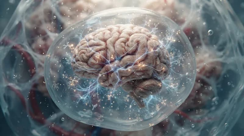
Cerebroids are 3D segments of ex vivo fetal brain tissue maintained in a highly controlled, nutrient-rich laboratory environment. By preserving the native structural integrity and cellular diversity of the dorsolateral prefrontal cortex, these models provide a far more faithful representation of the developing human brain than standard stem-cell-derived organoids. This advancement allows scientists to observe how the biological "scaffolding" of the cortex responds to external stressors in real-time, bridging the gap between basic cell culture and complex human development.
Decoding the Impact of IL-17A
A primary goal of the study was to investigate the biological pathways linking maternal immune activation—often triggered by infections during pregnancy—to potential neurodevelopmental risks in offspring. To do this, the team exposed their cerebroid models to IL-17A, a signaling protein associated with inflammatory immune responses. The results were immediate and structurally significant.
Microscopic analysis revealed that the presence of IL-17A caused the developing cortex to fold prematurely. Furthermore, the researchers observed an increase in cortical thickness alongside an accelerated timeline for neuron production and maturation. These physical shifts suggest that inflammatory signals do not merely stress the brain; they actively disrupt the precise, timing-dependent orchestration required for healthy cortical development.
Mechanisms of Action and Future Implications
By leveraging advanced RNA sequencing and proteomics, the team identified the NF-κB signaling pathway as a critical mediator in this process. When the researchers experimentally blocked this specific pathway, they successfully reversed many of the structural irregularities caused by the inflammatory protein. This discovery marks a significant step forward in understanding the cellular "gatekeepers" of fetal development.
Why It Matters
- Enhanced Modeling: Cerebroids offer superior accuracy compared to traditional organoids by retaining original tissue organization.
- Molecular Targeting: Identifying the NF-κB pathway as a focal point for inflammation provides a potential target for future protective strategies.
- Developmental Insights: The study helps demystify why maternal inflammation is statistically linked to neurodevelopmental conditions, moving the conversation from correlation to causal biological pathways.
Looking ahead, the research team plans to expand the use of the cerebroid system to other regions of the developing brain. By mapping how different specialized cells respond to inflammatory stimuli, scientists hope to eventually design models that can test potential interventions to shield the developing brain from environmental stressors, ultimately aiming to safeguard healthy cognitive development during gestation.